 Over the past several
years, media buzz and patient demand
have stimulated adoption of new surgical
devices and techniques before optimal
indications, patient selection, duration
of effect, and complications are well
known. The too-early adoption of new
techniques can lead to patient
disappointment and surgeon
dissatisfaction, resulting in early
abandonment of a procedure that
otherwise has great value, although
perhaps not for what it was originally
intended.
Over the past several
years, media buzz and patient demand
have stimulated adoption of new surgical
devices and techniques before optimal
indications, patient selection, duration
of effect, and complications are well
known. The too-early adoption of new
techniques can lead to patient
disappointment and surgeon
dissatisfaction, resulting in early
abandonment of a procedure that
otherwise has great value, although
perhaps not for what it was originally
intended.
Aesthetic surgeons are
practicing in a competitive commercial
environment in which patients are
eagerly looking for new technologies to
satisfy their desires for better,
quicker, and less invasive approaches
for facial rejuvenation. Product
development results in newer and better
devices, as evidenced by the progressive
development of Aptos sutures, Woffle
sutures, Contour sutures, Isse sutures,
and the new Silhouette suture approved
by the FDA in November 2006.
The Silhouette suture
shown here is stronger than conventional
barbed sutures, stands less chance of
breaking, and interacts with normal
tissues through surrounding collagen
tissue formation for better results. The
suture consists of a 25-cm-long 3-0
nonabsorbable polypropylene suture,
which is 50% thinner than the 2-0 used
in the design of the barbed sutures.
This decreases knot palpability and pain
in the temporal area. Suture strength is
maintained by avoiding oblique cuts in
the suture to create the "barbs" (30% to
50% loss of suture strength).
Each suture holds 11
clear, flexible, absorbable hollow cones
that allow for tissue growth inside and
around the cones, which are made from a
copolymer of glycolic acid and lactic
acid. An immediate anchoring effect is
created by the cones. A delayed
anchoring effect is produced by a series
of knots created on the distal 10 cm of
the suture, which is intercalated with
the cones.
The knots control the
position of the cones and provide
further anchoring of the soft tissue,
creating collagen deposition around and
inside each loop of the knot. These
sutures can easily and safely be removed
after the cones are completely absorbed
using a small microphlebectomy hook.
The copolymer cones
are soft, and avoid the pinprick effects
of the barbed suture. Their large size
(1.0 mm x 2.5 mm) creates a strong
grasping effect on the fibrous soft
tissue. The cones are hydrophilic,
resulting in absorption of water and a
softer feel. They generate more collagen
formation as the result of chemical
interaction with the tissues. The
physical design of the device creates
stronger soft-tissue support. The cones
are absorbed over 8 to 10 months; 50% of
the device volume is absorbed in about 5
months.
The sutures can be a
complement to brow-fixation and deep
midface devices.
|
Before & After |
 |
| Figure 1. A
closed midface Silhouette suture
suspension in a 62-year-old
patient, shown before and 4
months after surgery. |
The Closed Midface
Technique
A patient with the
following attributes is a good candidate
for the closed midface technique using
Silhouette sutures:
- has mild to
moderate midface ptosis;
- is in the
late-30s to mid-60s age range;
- wishes to avoid
scars associated with traditional
surgical lifts;
- accepts 1 week of
downtime;
- has realistic
expectations (accepts a modest lift,
a shorter period of improvement than
open techniques, and the need for
additional surgery or procedures to
maintain improvement or correct skin
laxity); and
- desires to
improve prior facial surgery.
Poor candidates for
this procedure are characterized by:
- excessive skin
laxity and wrinkling;
- very thin faces
(a moderate amount of facial fat is
needed to prevent suture
visibility);
- wide, heavy, or
fatty faces;
- thick, heavy,
sebaceous skin; and
- anticipation of
significant weight loss after
surgery.
The closed midface
technique is performed under local
anesthesia, with or without oral
sedation, and requires about 1 hour to
complete. Other complementary procedures
can be performed at the same time: for
example, botulinum toxin Type A (BTTA);
fillers for soft-tissue augmentation; or
liposuction of the jowl, jawline, and
neck.
An incision is made in
the temporal area approximately 2 to 3
cm behind the anterior hairline and is
carried down to the deep temporal fascia
(DTF), where a plane of blind
soft-tissue dissection is created
between the superficial temporal fascia
(STF) and DTF. This facilitates
deployment of the 6-inch-long straight
Silhouette suture needle into the
superficial fibrous adipose tissue of
the lower temporal area, midface, and
lower face.
To enhance scar-tissue
adherence in the midface, insert the
needles in pairs, passing the tip just
through the exit point in the skin.
Leave the needles in place, performing
subcutaneous undermining with a small
tissue dissector placed through the
temporal incision on each side of the
needles. Then pull the 6-inch needles
completely out of the skin. This will
leave intact bridges of tissue where
each suture has been placed.
|
Before & After |
 |
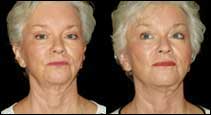
| Figure 2.
Endoscopic brow lift, endoscopic
subperiosteal midface lift with
Silhouette suture suspension,
neck lift, chin augmentation,
upper and lower eyelid lift, and
full-face laser resurfacing in a
48-year-old patient, shown
before and 4 months after
surgery. |
The needle exit points
are along a line drawn from the buccal
commissure medially to the gonion of the
mandible laterally. They begin just
lateral to the nasolabial fold and are
spaced 1 to 1.5 cm apart using a total
of four to six sutures per side
according to the patient's needs.
The curved taper
needles on the proximal end of the
suture are passed through the DTF and
then tied in pairs. Tissue repositioning
and suspension is accomplished first by
tying the knots in the temporal area;
further tissue movement and remodeling
are then achieved by placing tension on
the distal end of the suture and
massaging the tissues overlying the
sutures from medial to lateral.
Distal suture ends can
either be cut off at the skin at the
completion of the procedure or left long
to allow for further tissue shaping from
1 to 3 days after surgery. One-inch
adhesive closures or paper-tape strips
are used for 5 to 7 days to support the
facial tissues and minimize bruising and
swelling.
See Figure 1 for an
example of the closed midface procedure.
The Open Midface
Technique1,2
Good candidates for
the open technique are patients who
desire more dramatic, longer-lasting
midface elevation of the malar pads,
lower eyelids, or nasolabial folds, and
some more modest improvement of the
marionette area, jawline, and jaw. This
technique actually provides the combined
benefits of the open and closed methods.
Surgery is performed under general
anesthesia and requires about 2½ hours.
Other complementary procedures can be
performed at the same time, such as BTTA,
fillers, fat grafting, and liposuction.
Preoperatively, mark
the patient in the sitting upright
position, outlining the underlying bony
structures, the planned vectors of
correction, the path of the temporal
branch of the facial nerve, and the
superficial temporal artery. Initial
incisions and dissection are the same as
the closed technique.
Blind or illuminated
dissection with a periosteal elevator is
carried inferiorly, separating the STF
from the DTF down to the superior edge
of the zygomatic arch and medially to
the lateral orbital rim. For a modest
lift, sutures can be inserted as
previously described.
For a more dramatic
lift, subperiosteal dissection over the
middle and medial zygoma and maxilla is
completed using illuminated direct
vision or the endoscope. An optional
1.5-cm incision can be made in the
buccal sulcus bilaterally to make
subperiosteal dissection of the maxilla
and medial zygoma easier to complete
with headlight or retractor
illumination.
|
Before & After |
 |
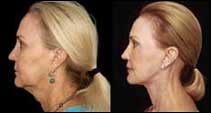 |
| Figure 3.
Subperiosteal midface lift with
Silhouette sutures; neck lift;
fat grafting to the nasolabial
folds, marionette lines, and
upper and lower lips; laser
resurfacing of the forehead,
lateral canthal area, and upper
and lower eyelids in a
65-year-old patient, shown
before and 4 months after
surgery. |
Insertion of two to
four sutures per side may be done
blindly or with illumination. Each
needle is inserted in the space created
between the STF and DTF down over the
zygoma in the subperiosteal space.
Sinusoidal advancement of the long,
straight 6-inch needle is made through
the malar pad and soft tissue of the
cheek, exiting the skin past the malar
pad and lateral to the nasolabial fold
along the points described in the
closed-suture procedure.
The proximal taper
needles are passed through the DTF in
the temporal area and tied in pairs.
While applying traction on the distal
end of the Silhouette suture, move the
cheek tissues superiorly and laterally,
allowing the cones to engage several
layers of tissue to create volumetric
stacking of soft tissue. Sutures may be
clipped at the skin at the completion of
surgery or left long for sequential
soft-tissue molding during the 1 to 3
days after surgery.
Slight overcorrection
is suggested to counter soft-tissue
settling during the 1 to 3 months after
surgery. Adhesive closures or strips of
paper tape support the soft tissue for 5
to 7 days. This technique simplifies
midface suspension with the added
benefit of postoperative adjustment.
See Figure 2 (page 46)
and Figure 3 (this page) for clinical
examples of the open technique.
In summary, the new
Silhouette suture offers improved
results with closed and open midface
rejuvenation. It can be used alone or in
combination with other internal suture
or fixation devices.
N.D. Moscoe, MD,
FACS, is a board-certified plastic
surgeon in private practice in Austin,
Tex. He is affiliated with all major
Austin hospitals, including Bailey
Square Surgery Center, Brackenridge
Hospital, and St David's Hospital. He
can be reached at ndmoscoe@sbcglobal.net
or via his Web site,
www.ndmoscoemd.com.
Nicanor Isse, MD,
is a board-certified plastic surgeon and
the founder and president of the Isse
Institute of Cosmetic Surgery in Newport
Beach and Burbank, Calif. He is
affiliated with four hospitals in
Southern California and three
universities in California and Italy. He
can be reached at (818) 557-6595 or
drissenewport@sbcglobal.net.
References
- Paul MD. Using
barbed sutures in open/subperiosteal
midface lifting. Aesthetic Surg J.
2006; 26:725–732.
- Hobar PC, Flood
J. Subperiosteal rejuvenation of the
midface and periorbital area: A
simplified approach. Plast
Reconstr Surg. 1999;104:
842–851.